2026-09-11
Diagnosed with Diabetes? Start with an Eye Exam — Even with Clear Vision, Your Retina May Already Be Damaged
Even with 20/20 vision, diabetic retinopathy can silently damage the retina. Learn screening schedules, disease stages, macular edema, and modern treatments.


First, let me reassure you.
Having diabetes does not necessarily mean your vision will deteriorate.
In fact, the vast majority of individuals diagnosed with diabetic retinopathy are still in the early stages, where active medical treatment is often unnecessary—careful blood sugar management and regular observation are usually sufficient.
However, there is a critical distinction: diabetic retinopathy is a disease where the strategy of "visiting the clinic only when symptoms appear" does not work.
This is because vision decline typically occurs only after a significant portion of the retina has already sustained severe damage.
Therefore, for this disease in particular, you must schedule your examinations according to the calendar, not according to symptoms.
The guidelines are clear and unequivocal:
For type 2 diabetes, you should receive your first ophthalmologic evaluation on the day of diagnosis; for type 1 diabetes, within 5 years of diagnosis. Thereafter, annual screening is recommended at least once a year
(Korea Disease Control and Prevention Agency, National Health Information Portal).
Yet, what is the reality?
A study analyzing 3,717 diabetic patients aged 40 and older using data from the Korea National Health and Nutrition Examination Survey (2016–2021) revealed that only 1,109 individuals—29.5%—had undergone a fundus examination within the preceding year.
Furthermore, this screening rate did not show meaningful improvement over the 6-year period
(Kim MS, Park SJ, Joo K, Woo SJ, Journal of Korean Medical Science, 2024).

Hello, this is St. Mary's Jin Eye Clinic.
Today, we will detail what diabetic retinopathy actually does within the eye, when and how often you need to be screened, and what modern treatments can—and cannot—achieve, based strictly on clinical evidence.
[Table of Contents]
What exactly is diabetic retinopathy?
Why are there no symptoms in the early stages?
When and how often should examinations be performed?
What tests are conducted at the clinic?
How are the stages of the disease classified?
What is diabetic macular edema?
Does strict blood sugar control truly reduce ocular damage?
Is it true that rapid glycemic control can worsen eye conditions?
How is it treated? Can lost vision be restored?
Red flags requiring immediate, same-day medical attention
Other ocular complications caused by diabetes
Frequently Asked Questions
1. What exactly is diabetic retinopathy?
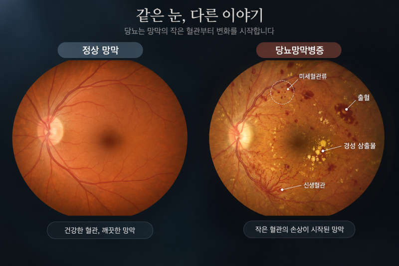
The retina is a delicate, thin neural layer lining the inside of the eye.
Analogous to the film in a traditional camera, this neural layer receives oxygen and nutrients through a complex microvascular network.
When blood glucose levels remain chronically elevated, these fine capillaries sustain damage first. The progression typically unfolds in the following sequence:
Capillary walls weaken, giving rise to tiny outpouchings known as microaneurysms.
Blood components leak through these compromised vessel walls. Dot-and-blot hemorrhages and hard exudates (bright lipid deposits) become visible on fundus photography.
Certain microvessels become completely occluded. The affected regions of the retina suffer from chronic ischemia (oxygen deprivation).
Oxygen-deprived retinal tissue secretes signaling molecules, primarily vascular endothelial growth factor (VEGF), demanding the formation of new blood vessels.
Prompted by these signals, fragile neovascular vessels proliferate. Because these vessels are hastily formed, their walls are exceptionally fragile and prone to rupture.
Once this stage is reached, the severity of the pathology escalates dramatically.
When these neovascular vessels rupture, vitreous hemorrhage occurs, causing sudden vision obstruction. As fibrovascular tissue proliferates along these abnormal vessels, it exerts traction on the retina, leading to tractional retinal detachment.
Furthermore, if neovascularization invades the anterior chamber angle—the drainage pathway for aqueous humor—it precipitates neovascular glaucoma, a condition notoriously refractory to treatment.
In essence, diabetic retinopathy is not merely a localized ocular disorder; rather, it represents the ocular manifestation of systemic microvascular damage.
The retina is the only site in the human body where living vascular networks can be visualized non-invasively and directly.
Consequently, a single fundus photograph provides an invaluable window into the systemic microvascular health of the entire body.
2. Why are there no symptoms in the early stages?
This question touches upon the most critical aspect of the disease.
First, the retina lacks pain sensory fibers. Retinal damage causes no somatic pain or physical discomfort.
Second, early lesions predominantly arise in the peripheral retina.
Our high-acuity central vision is managed almost entirely by the macula, a minute region at the very center of the retina. Even if dozens of microaneurysms develop in the peripheral retina, visual acuity charts can still read 20/20 (1.0) as long as the macula remains unaffected.
Third, binocular compensation masks unilateral deficits.
When visual deterioration begins in one eye, the contralateral eye compensates, making the impairment unnoticeable until the patient inadvertently covers one eye.
As a result, clinical encounters often reveal striking disparities:
A patient presenting with 20/20 visual acuity may show multiple retinal hemorrhages and hard exudates on fundus imaging, whereas a patient who finally presents complaining of "mild blurring" has often already developed diabetic macular edema or vitreous hemorrhage.
In diabetic retinopathy, subjective visual acuity is the last indicator to show decline.
Data from the Korea Disease Control and Prevention Agency confirm this discrepancy:
According to the 2017 Korea National Health and Nutrition Examination Survey, the prevalence of diabetic retinopathy among diabetic patients aged 40 and older was 19.6%, yet only 23.5% had undergone a fundus examination to detect complications
(KDCA National Health Information Portal).
Without proactive screening, the disease progresses entirely unnoticed.
The domestic disease burden is also climbing steadily.
An epidemiological study utilizing nationwide health survey data showed that the prevalence of diabetes among adults aged 40 and older rose from 13.2% in 2008 to 17.3% (approximately 4.98 million individuals) in 2020. Concurrently, the prevalence of diabetic retinopathy among diabetic individuals rose from 16.6% to 24.6% (approximately 1.23 million individuals).
The same study projected that by 2040, the prevalence of diabetic retinopathy could reach 38.8%, representing roughly 3.15 million patients (Kim MS, Nam S, Lee J, Woo SJ, Journal of Korean Medical Science, 2026).
3. When and how often should examinations be performed?
Classification | Initial Exam Timing | Subsequent Interval |
Type 2 Diabetes | Day of diagnosis | At least once a year |
Type 1 Diabetes | Within 5 years of diagnosis | At least once a year |
Pre-pregnancy & Pregnancy | Prior to conception & early 1st trimester | Every 3 months minimum |
Existing Retinopathy | Immediately | Every 3–6 months (by stage) |
(KDCA National Health Information Portal / American Academy of Ophthalmology, Diabetic Retinopathy Preferred Practice Pattern)
Why is an immediate exam required on the day of diagnosis for Type 2 Diabetes?
Because type 2 diabetes develops insidiously, chronic hyperglycemia has frequently been present for years prior to formal clinical diagnosis.
Consequently, a notable proportion of patients already present with established diabetic retinopathy on the day they are first diagnosed.
In contrast, type 1 diabetes exhibits an acute clinical onset, permitting a 5-year monitoring grace period.
Why are evaluations required every 3 months during pregnancy?
Profound hormonal fluctuations and systemic hemodynamic shifts during pregnancy can accelerate retinopathy progression at an unusually rapid rate.
It is therefore prudent to assess baseline retinal status prior to conception.
However, gestational diabetes diagnosed de novo during pregnancy does not warrant this specific retinopathy screening protocol
(AAO Preferred Practice Pattern).
This heightened surveillance applies specifically to women with preexisting diabetes prior to pregnancy.
"I had an exam last year and was told everything was fine" — this does not guarantee stability this year.
The 1-year screening interval exists not because one is considered "safe for a full year," but because significant vascular changes can readily develop within that 12-month span.
4. What tests are conducted at the clinic?
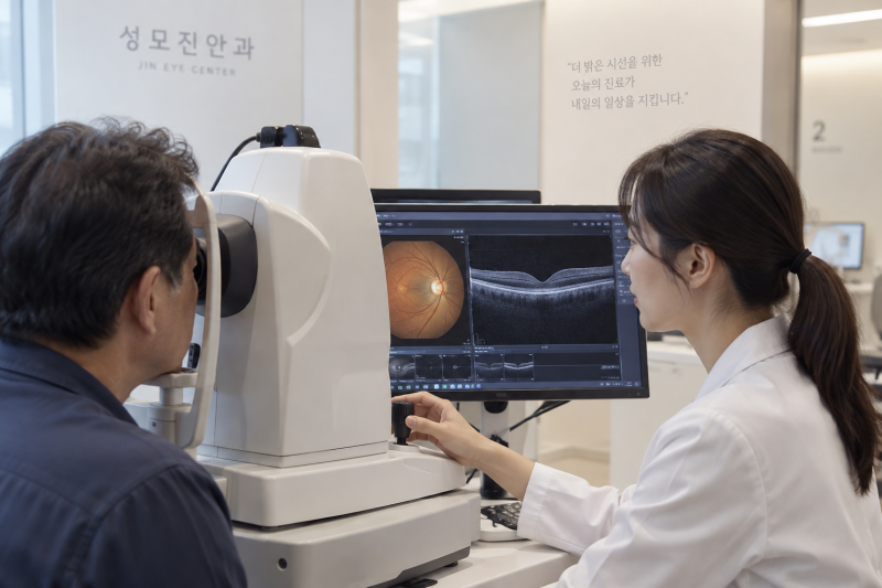
① Visual Acuity & Refraction Assessment — Establishes a baseline for best-corrected visual acuity.
Having precise numerical baselines is essential for comparative monitoring across subsequent visits.
② Dilated Fundus Examination — The definitive core of diabetic eye disease evaluation.
Following pharmacologic pupillary dilation (mydriasis), the entire retinal expanse is directly inspected.
The clinical stage is graded by evaluating microaneurysms, intraretinal hemorrhages, hard exudates, areas of capillary nonperfusion, and neovascularization.
③ Optical Coherence Tomography (OCT) — Delivers micron-level cross-sectional imaging of the macula.
This non-contact, completely painless scan takes only seconds.
It accurately detects macular edema—subretinal or intraretinal fluid accumulation—that cannot be reliably evaluated on two-dimensional fundus photography alone. It captures subclinical edema before visual acuity declines.
④ OCT Angiography (OCTA) — Evaluates segmented retinal microvascular layers without contrast injection.
It assesses the expansion of the foveal avascular zone (FAZ), areas of nonperfusion, and early neovascular buds.
⑤ Fluorescein Angiography (FA) — When indicated, intravenous contrast dye is administered through an arm vein to visualize precise leakage points and capillary dropouts.
This is especially valuable when determining the extent of targeted retinal photocoagulation.
Our clinic is equipped with advanced OCT and OCTA platforms, enabling comprehensive structural and vascular assessment through simultaneous fundus and macular evaluation.
Helpful Instructions Prior to Your Visit:
Mydriatic eye drops dilate the pupils, causing photophobia (glare sensitivity) and blurred near vision for approximately 4 to 6 hours.
Please refrain from driving yourself on the examination day, and bringing sunglasses is highly recommended for comfort.
Because pupillary dilation requires adequate time to take effect, please allow sufficient time for your appointment.
Take all prescribed diabetes medications and insulin as scheduled.
Fasting is not required for a fundus examination.
In fact, prolonged fasting during clinical visits poses a risk of hypoglycemia; arriving having eaten normally is much safer.
Knowing your most recent glycated hemoglobin (HbA1c) level and the date of your initial diabetes diagnosis helps us evaluate your condition with greater precision.
5. How are the stages of the disease classified?
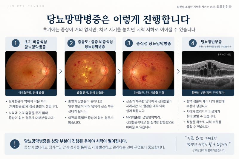
Stage | Pathologic Events Inside the Eye | Typical Patient Symptoms |
Non-Proliferative (Early) | Emergence of sporadic microaneurysms and dot hemorrhages | Virtually absent |
Non-Proliferative (Moderate–Severe) | Expansion of areas with occluded capillary blood flow | Virtually absent |
Proliferative | Neovascular growth leading to hemorrhage and retinal detachment | Sudden visual field obstruction |
Diabetic Macular Edema | Fluid accumulation causing macular thickening (can arise at any stage) | Blurred text, straight lines appearing wavy |
Please pay close attention to the middle two rows of this table.
Across non-proliferative diabetic retinopathy—whether mild, moderate, or severe—subjective symptoms remain virtually nonexistent.
The fundamental takeaway here is that "absence of symptoms" is by no means synonymous with "absence of pathology."
Furthermore, diabetic macular edema can develop independently of retinopathy severity.
A patient with early non-proliferative retinopathy may suffer pronounced vision loss due to macular edema, while another with advanced proliferative retinopathy might retain 20/20 vision because the macula remains anatomically preserved.
This is why fundus photography and OCT cross-sectional imaging must be evaluated together.
6. What is diabetic macular edema?
Diabetic macular edema is the leading cause of moderate to severe vision loss in diabetic retinopathy.
When fluid leaks from damaged, permeable microvessels into the macula, the retinal layers become swollen and thickened.
This is structurally comparable to a photograph film becoming warped and distorted from water damage.
Consequently, fine text blurs, straight lines appear distorted or wavy (metamorphopsia), and contrast sensitivity drops.
A telltale characteristic is persistent blurring that cannot be corrected with a new eyeglass prescription.
Proliferative retinopathy tends to compromise vision abruptly, whereas macular edema degrades vision gradually.
Because sudden visual loss triggers immediate alarm, patients promptly seek emergency care; however, insidious, gradual blurring is frequently dismissed as "normal aging or presbyopia," resulting in critical therapeutic delays.
Distortion of straight lines also occurs in age-related macular degeneration. Because the underlying etiology and clinical management differ entirely, this symptom must be accurately evaluated with diagnostic imaging rather than self-assessed.
7. Does strict blood sugar control truly reduce ocular damage?
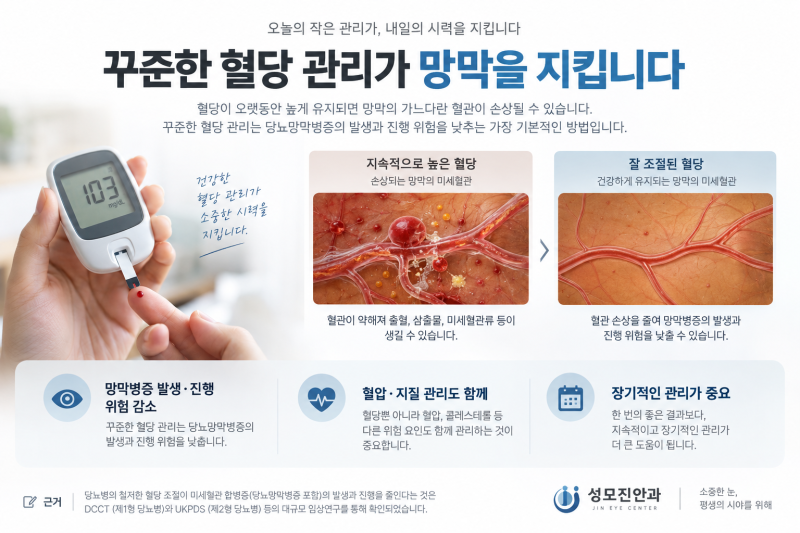
Yes. This is backed by robust, high-level clinical evidence.
In Type 1 Diabetes:
In the landmark Diabetes Control and Complications Trial (DCCT) enrolling 1,441 patients, the intensive therapy group maintained blood glucose levels near normal ranges over a mean follow-up of 6.5 years, reducing the risk of developing or worsening retinopathy by up to 76% compared to the conventional treatment group
(DCCT Research Group, New England Journal of Medicine, 1993 / DCCT/EDIC Research Group, Diabetes, 2015).
In Type 2 Diabetes:
In the UKPDS 33 trial involving 3,867 participants, the intensive treatment cohort achieved a median HbA1c of 7.0% compared to 7.9% in the conventional group.
This yielded a 25% reduction in microvascular complications (RR 0.75, 95% CI 0.60–0.93), including a 29% risk reduction in the requirement for retinal laser photocoagulation (RR 0.71, 95% CI 0.53–0.96)
(UK Prospective Diabetes Study Group, The Lancet, 1998).
Two essential clinical realities must be highlighted alongside these findings:
First, the ocular benefits of glycemic control manifest gradually over several years.
Lowering your HbA1c by 0.5% this month will not produce immediate visible clearing of retinal lesions next month. Viewed from another angle, rigorous blood sugar control today is an investment protecting your vision 5 to 10 years down the line.
Second, blood glucose is not the sole determinant.
Strict control of blood pressure, management of dyslipidemia, and complete smoking cessation must occur concurrently. Furthermore, declining renal function demands immediate retinal evaluation, as microvascular pathology damages the renal glomeruli and retinal capillary beds through identical pathophysiology.
8. Is it true that rapid glycemic control can worsen eye conditions?
This concern is neither entirely baseless nor a valid justification for delaying glycemic control.
Because this question arises frequently, let us review the exact clinical evidence.
Secondary analyses of the aforementioned DCCT trial clarify this phenomenon:
Among 1,439 patients (711 intensive therapy, 728 conventional therapy), early transient worsening of retinopathy occurred in 13.1% of the intensive group compared to 7.6% of the conventional group.
Rapid glycemic reduction can indeed induce a temporary early worsening of retinal pathology.
However, the long-term clinical trajectory is what truly matters:
By 18 months, 51% of affected eyes in the intensive cohort and 55% in the conventional cohort had completely recovered to baseline status, and not a single patient experienced severe, irreversible vision loss as a consequence of this early worsening.
The study authors concluded that "the long-term benefits of intensive insulin therapy vastly outweigh the transient risks of early worsening"
(DCCT Research Group, Archives of Ophthalmology, 1998).
Therefore, clinical management should follow these practical principles:
▶ Never delay optimal glycemic control; long-term clinical benefit is indisputable.
▶ Always establish your baseline retinal status via fundus examination prior to initiating aggressive glucose-lowering regimens. If advanced retinopathy is present, titration speed and ophthalmic follow-up intervals can be adjusted collaboratively.
▶ Patients with prolonged, poorly controlled diabetes who are newly starting insulin therapy, or those experiencing rapid glucose reductions following metabolic/bariatric surgery, warrant more frequent ophthalmic monitoring during the first 12 months.
Lowering blood glucose is not hazardous in itself; lowering it without knowing the underlying state of your retina is where the clinical risk lies.
9. How is it treated? Can lost vision be restored?

Modality | Mechanism of Action | Clinical Limitations |
Panretinal Photocoagulation (Laser) | Ablates ischemic peripheral retina to suppress neovascular drives | Possible peripheral visual field and night vision reduction |
Intravitreal Injections (Anti-VEGF) | Seals hyperpermeable vessels to resolve macular edema | Requires repeated injections; rare risk of endophthalmitis |
Pars Plana Vitrectomy (Surgery) | Clears vitreous hemorrhage/fibrovascular membranes and reattaches retina | Postoperative cataract progression, risk of rebleeding |
① Panretinal Photocoagulation (PRP Laser)
This is the gold-standard treatment for proliferative diabetic retinopathy.
Applying laser burns to the ischemic peripheral retina reduces overall retinal oxygen demand and suppresses intraocular VEGF production, thereby arresting neovascular growth. Though an established modality for decades, its therapeutic efficacy is thoroughly proven.
In the landmark Diabetic Retinopathy Study (DRS), severe visual loss at 2 years occurred in 16.3% of untreated control eyes compared to only 6.4% of eyes treated with panretinal laser photocoagulation.
However, this therapy involves an unavoidable functional tradeoff:
Because peripheral retinal tissue is deliberately ablated, patients may experience peripheral visual field constriction and reduced night vision (nyctalopia).
The therapeutic objective of PRP is not to enhance baseline acuity, but rather to sacrifice peripheral retina in order to preserve vital central vision.
② Intravitreal Anti-VEGF Injections
This is the first-line pharmacotherapy for diabetic macular edema.
Following local topical anesthesia, medication is delivered into the vitreous cavity using an ultrafine needle. The injection procedure itself takes only moments.
Clinical efficacy has been extensively validated. In a head-to-head randomized trial evaluating three anti-VEGF agents in 660 patients, mean visual acuity improved by 10.0 to 12.8 letters at 2 years; eyes with worse baseline visual acuity (20/50 or worse) demonstrated even greater improvements of 13.3 to 18.1 letters
(Wells JA et al., Ophthalmology, 2016; DRCR.net Protocol T).
Anti-VEGF therapy can also serve as an effective alternative to PRP in proliferative diabetic retinopathy.
At 5-year follow-up, visual acuity outcomes were comparable between both modalities (mean 20/25 in both arms), but the injection group demonstrated less visual field loss (cumulative -330 dB vs. -527 dB), a lower rate of vitrectomy (11% vs. 19%), and reduced development of vision-impairing macular edema (22% vs. 38%)
(Gross JG et al., JAMA Ophthalmology, 2018; DRCR.net Protocol S).
Nonetheless, anti-VEGF therapy comes with distinct considerations:
It requires periodic, ongoing injections over several years.
Premature discontinuation can lead to recurrent edema and rapid visual relapse; indeed, the cited study identified patient compliance and treatment adherence as pivotal criteria when selecting injection monotherapy.
Common mild adverse events include subconjunctival hemorrhage, transient ocular soreness, and temporary floaters, while rare serious complications include infectious endophthalmitis, intraocular pressure elevation, and retinal detachment.
③ Pars Plana Vitrectomy
Surgical intervention is indicated for non-clearing vitreous hemorrhage or tractional retinal detachment secondary to fibrovascular proliferation.
The vitreous blood and proliferative membranes are micro-surgically excised, and the detached retina is anatomically reapposed.
Postoperative risks include accelerated nuclear cataract progression, recurrent vitreous hemorrhage, and potential retinal redetachment.
Focal/grid laser photocoagulation is also utilized for macular edema.
The Early Treatment Diabetic Retinopathy Study (ETDRS) demonstrated that focal macular laser reduced the 3-year risk of moderate visual loss by approximately 50%
(ETDRS Report No. 1, Archives of Ophthalmology, 1985).
Today, intravitreal anti-VEGF agents represent the primary therapy, while focal laser is utilized either in combination or selectively depending on the lesion’s precise anatomic location.
Four critical therapeutic principles must always be understood:
First, these interventions do not eradicate the underlying disease; they arrest or slow its progression.
Second, necrotic retinal neural tissue cannot be regenerated. In chronic macular edema where neuroretinal layers have sustained structural thinning, or in long-standing tractional detachments, eliminating fluid and flattening the retina cannot fully restore baseline visual acuity.
Third, the timing of intervention determines the ultimate functional prognosis. While eyes with poorer baseline vision in injection trials showed larger letter gains, their final absolute visual acuity rarely equaled that of eyes treated before vision deteriorated.
Fourth, undergoing medical treatment does not eliminate the need for ongoing screening. Even after successful procedures, systemic diabetes persists, and retinal microvascular changes continue to evolve.
10. Red flags requiring immediate, same-day medical attention

Although we emphasize not waiting for symptoms to appear, any onset of the following acute manifestations demands same-day ophthalmologic assessment:
Sudden appearance of a dark curtain or shadow obscuring part of the visual field — Suggestive of massive vitreous hemorrhage or retinal detachment.
A sudden shower of floaters, cobwebs, or dark spots — Especially if developing over hours, which often heralds active intraocular bleeding.
Acute blurring or loss of vision in one eye — Indicative of macular edema, acute hemorrhage, or retinal vascular occlusion.
Severe eye pain accompanied by ciliary injection (redness) and reduced vision — Suggestive of acute intraocular pressure elevation due to neovascular glaucoma.
These four presentations must never be postponed until your next scheduled check-up. Proliferative diabetic retinopathy can escalate severely within days.
11. Other ocular complications caused by diabetes
Diabetes affects structures throughout the entire ocular system, not just the retina:
▶ Cataracts develop at an earlier age and progress more rapidly.
Accumulation of sorbitol and osmotic stress inside the crystalline lens accelerates cataractous opacification. Consequently, diabetic patients frequently require cataract surgery at significantly younger ages than non-diabetic peers.
▶ Rapid glycemic fluctuations cause acute refractive shifts over days.
Osmotic changes alter the hydration and curvature of the crystalline lens, changing refractive error. Updating eyeglasses while blood sugar is fluctuating produces prescriptions that quickly become inaccurate. New lenses should be prescribed only after glycemic stabilization.
▶ Neovascular Glaucoma — As previously described, unchecked proliferative retinopathy stimulates abnormal neovascular membranes across the trabecular meshwork, producing catastrophic intraocular pressure spikes.
▶ Dry Eye Syndrome & Delayed Corneal Epithelial Healing — Chronic diabetic neuropathy reduces corneal sensation and impairs epithelial barrier repair, complicating contact lens wear and slowing recovery following ocular surgeries.
▶ Ocular Motor Cranial Nerve Palsies (Diabetic Neuropathy) — Microvascular ischemia to cranial nerves (CN III, IV, or VI) can cause sudden-onset binocular diplopia (double vision) or ptosis (eyelid drooping). Although these usually resolve spontaneously over several months, urgent neuro-ophthalmic evaluation is essential to rule out intracranial aneurysms or stroke.
A practical tip for holiday gatherings with elderly parents:
Take a moment to ask these three simple questions:
"How many years have you been taking your diabetes medication?"
"When was the last time you had a dilated fundus photo taken of the inside of your eyes?"
"How clear does small newspaper print look to you these days?"
You may be surprised by how often the answer to the second question is, "I have never had that done."
Family gatherings offer an ideal opportunity to review the ocular health of your loved ones.
Pediatric visual development and myopia management were covered comprehensively in our previous column.
12. Frequently Asked Questions
Q. Should I still get an eye exam if my visual acuity is 20/20 (1.0)?
Yes, absolutely.
Visual acuity reflects only the tiny central macula, whereas diabetic retinopathy typically starts in the peripheral retina.
It is exceptionally common in ophthalmic practice to observe multiple hemorrhages and microaneurysms on fundus photography in patients with 20/20 vision.
A standard vision test cannot substitute for a fundus examination.
Q. Can I skip retinal screening if my HbA1c is well-controlled?
No. Good glycemic control reduces risk significantly, but it does not eliminate it entirely.
Furthermore, screening frequency is determined primarily by the total duration of diabetes and current retinal findings, rather than a single glycemic reading.
If your blood sugar is under excellent control, your next exam simply provides positive confirmation of ocular stability.
Q. Am I unable to drive after a fundus examination?
Mydriatic drops keep the pupils enlarged for 4 to 6 hours, producing considerable light sensitivity and near-focus blur.
Driving yourself home in this condition is unsafe and strictly discouraged.
Using public transit or having a companion drive is advisable. Bringing a pair of sunglasses will make your return home significantly more comfortable.
Q. Is the examination painful?
Standard fundus photography and OCT imaging are entirely non-contact and painless.
Mydriatic eye drops may cause a brief, mild stinging sensation upon instillation.
If diagnostic fluorescein angiography is required, an intravenous line in the arm is necessary, and urine may appear bright yellow for several hours following the test.
Q. Does retinal laser photocoagulation improve visual acuity?
No. Panretinal photocoagulation is designed to prevent further deterioration; in fact, it often causes some loss of peripheral visual field and night vision.
Ophthalmologists recommend laser because accepting these mild functional tradeoffs is far preferable to risking irreversible loss of central vision.
This risk-benefit profile is thoroughly discussed with patients prior to treatment.
Q. Once intravitreal eye injections are started, must they be continued for life?
While "lifelong" is not necessarily the case, single-dose resolution is exceedingly rare.
Treatment typically begins with monthly loading doses, gradually extending the treatment interval based on anatomical response.
Injections may be paused if macular dry status stabilizes, though vigilant clinical monitoring remains mandatory.
Q. Can patients with diabetic retinopathy undergo refractive surgeries like LASIK or LASEK?
This depends entirely on individual retinal stability and systemic glycemic control.
Refractive surgery is contraindicated in the presence of active, progressing retinopathy or unstable blood glucose.
Although refractive surgery operates on the cornea, diabetes affects corneal nerve regeneration and surface wound healing; a comprehensive evaluation of both corneal and retinal health is required before determining candidacy.
Q. I recently had fundus photos taken during a general health check-up. Do I still need an ophthalmology visit?
A general health check-up photo is a great starting point.
However, screening health check-ups typically capture only one or two non-mydriatic images, which cannot sufficiently evaluate the peripheral retina or provide micron-level macular cross-sections.
If you bring your health check-up result sheets, we will review the initial images and determine whether supplemental testing is warranted.
For more in-depth clinical discussions on eye health, visit the St. Mary's Jin Eye Clinic YouTube channel, 'Jjin Eye'.
Conclusion
Please keep these three key facts in mind:
First, schedule screening based on the calendar, not on symptoms.
For type 2 diabetes, screen on the day of diagnosis; for type 1, within 5 years; thereafter, at least once annually.
Second, good visual acuity merely indicates that the macula is not yet compromised.
The health of the peripheral retina can only be confirmed through a direct fundus evaluation.
Third, retinal treatments are designed to halt progression, not restore lost tissue.
Therefore, when you initiate treatment dictates your ultimate visual prognosis.
When caught in the early stages, diabetic retinopathy is highly manageable, often requiring nothing more than routine observation and lifestyle control.
The most dependable protection you can provide for your future vision is a single fundus photograph—taken before any symptoms ever appear.
Please feel free to reach out if you have any questions.
St. Mary's Jin Eye Clinic
337 Gangnam-daero, Seocho-gu, Seoul (1329-10 Seocho-dong)
5-minute walk from Gangnam Station Exit 5 · Free parking available within building
Clinic Hours
Weekdays: 09:00 – 18:00 (Lunch 13:00 – 14:00)
Saturdays: 09:00 – 15:00 (No lunch break)
Closed on Sundays and Public Holidays
Phone: 02-577-7782
※ This article is provided for general informational and educational purposes only and should not replace individual clinical diagnosis or medical treatment.
Quoted clinical trial statistics reflect specific study cohorts; actual individual outcomes may vary depending on diabetes duration, glycemic stability, systemic comorbidities, and concurrent medications.
All medical and surgical interventions carry inherent limitations and risks. Diagnostic and therapeutic decisions should always be made in consultation with an ophthalmologist and your primary managing physician.
[References]
Kim MS, Park SJ, Joo K, Woo SJ. Trends and Barriers in Diabetic Retinopathy Screening: Korea National Health and Nutritional Examination Survey 2016–2021. Journal of Korean Medical Science. 2024;39(27):e203
Kim MS, Nam S, Lee J, Woo SJ. Nationwide Trends and Future Projections of Diabetes and Diabetic Retinopathy Prevalence in Korea: Korean National Health and Nutrition Examination Survey Study. Journal of Korean Medical Science. 2026;41(3):e31
Korea Disease Control and Prevention Agency National Health Information Portal. Diabetic Retinopathy.
American Academy of Ophthalmology. Diabetic Retinopathy Preferred Practice Pattern®. Ophthalmology.
The Diabetes Control and Complications Trial Research Group. The effect of intensive treatment of diabetes on the development and progression of long-term complications in insulin-dependent diabetes mellitus. New England Journal of Medicine. 1993;329(14):977-986
DCCT/EDIC Research Group. Effect of intensive diabetes therapy on the progression of diabetic retinopathy in patients with type 1 diabetes: 18 years of follow-up in the DCCT/EDIC. Diabetes. 2015;64(2):631-642
The Diabetes Control and Complications Trial Research Group. Early worsening of diabetic retinopathy in the Diabetes Control and Complications Trial. Archives of Ophthalmology. 1998;116(7):874-886
UK Prospective Diabetes Study (UKPDS) Group. Intensive blood-glucose control with sulphonylureas or insulin compared with conventional treatment and risk of complications in patients with type 2 diabetes (UKPDS 33). The Lancet. 1998;352(9131):837-853
Early Treatment Diabetic Retinopathy Study Research Group. Photocoagulation for diabetic macular edema: ETDRS Report Number 1. Archives of Ophthalmology. 1985;103(12):1796-1806
The Diabetic Retinopathy Study Research Group. Photocoagulation treatment of proliferative diabetic retinopathy. (DRS Reports)
Wells JA, Glassman AR, Ayala AR, et al. Aflibercept, Bevacizumab, or Ranibizumab for Diabetic Macular Edema: Two-Year Results from a Comparative Effectiveness Randomized Clinical Trial. Ophthalmology. 2016;123(6):1351-1359
Gross JG, Glassman AR, Liu D, et al. Five-Year Outcomes of Panretinal Photocoagulation vs Intravitreous Ranibizumab for Proliferative Diabetic Retinopathy: A Randomized Clinical Trial. JAMA Ophthalmology. 2018;136(10):1138-1148