2026-09-09
If Door Frames or Tile Grout Lines Look Distorted — Check at Home in 30 Seconds, One Eye at a Time
Discover how to spot early signs of macular degeneration using the Amsler grid at home, understand dry vs. wet AMD, and learn why prompt testing is vital for vision preservation in older adults.


First, let us reassure you about one thing.
Slightly distorted or wavy lines can occur when your eyes are fatigued or when your tear film is dry; in most cases, it is not a serious condition.
However, there is one critical catch.
If you cover one eye and straight lines still appear distorted, or if the center of a letter looks blank or missing, the story completely changes.
This is a warning sign of a problem in the macula—the part of the eye responsible for central vision. Among macular conditions, wet (exudative) age-related macular degeneration (AMD) is a disease where the timing of treatment initiation determines your final visual outcome.
Let us look at the numbers to see how serious this is.
In a study analyzing fundus photographs of 13,737 individuals aged 40 and older from the Korea National Health and Nutrition Examination Survey (2017–2020), 2,222 participants (16.2%) showed signs of macular degeneration.
Shockingly, 95.25% of them had no idea they had the condition
(Kim Y, Han K, Kim JH, Scientific Reports, 2025).
While most were still in the early stages, what this number tells us is clear:
Macular degeneration begins quietly without symptoms, and by the time you actually notice visual changes, it has often already progressed.
Hello, this is St. Mary's Jin Eye Clinic.
Today, we will cover what macular degeneration is, how to check for it at home using an Amsler grid, and explain precisely what this self-test can and cannot detect based on clinical evidence.
[Table of Contents]
Where is the macula, and why is it important?
What does vision look like with macular degeneration?
How do you use the Amsler grid?
Can you rely solely on the Amsler grid?
Dry vs. Wet: What is the difference?
What is my personal risk level?
What tests are performed at the clinic?
How is it treated? Can lost vision be restored?
Do supplements (Lutein / AREDS2) really help?
What lifestyle changes can lower my risk?
It might not be macular degeneration
Frequently Asked Questions
1. Where is the macula, and why is it important?
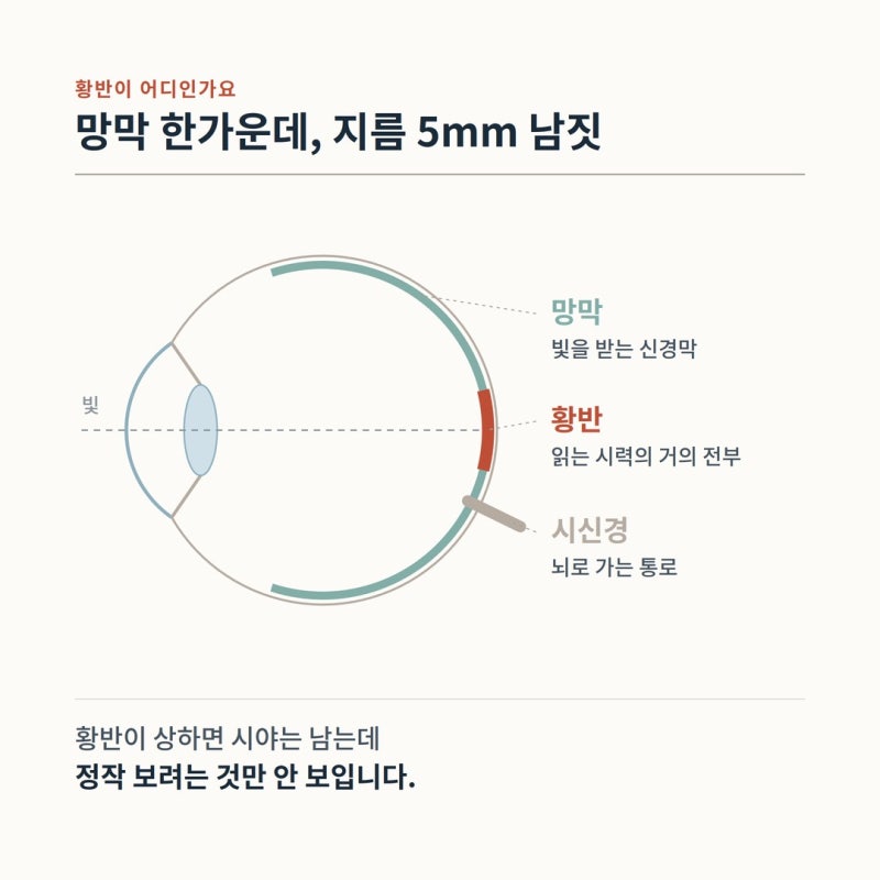
The thin layer of nerve tissue lining the back of the eye is the retina.
In camera terms, it functions like the film or digital sensor.
Right at the center of this retina—the exact tiny focal point where your line of sight lands—lies the macula.
It is only about 5 mm in diameter, and its most sensitive center (the fovea) measures less than 1 mm.
Yet, this microscopic area carries out an overwhelming share of your visual work:
▶ Reading vision—virtually all of what we consider 20/20 vision
▶ The ability to distinguish colors
▶ The ability to recognize faces
The retina outside the macula is responsible for peripheral field of vision.
Therefore, when the macula is damaged, you develop a very distinct disability: your peripheral view remains intact, but you cannot see what you are directly focusing on.
You can walk around without bumping into walls, but you cannot read the newspaper, and the center of the face of the person sitting across from you appears blurred or erased.
Age-related macular degeneration (AMD) is a disease in which this macula degenerates with age.
It begins as cellular waste products (drusen) accumulate beneath the retina, and the retinal pigment epithelium and choroid that nourish the macula begin to deteriorate.
2. What does vision look like with macular degeneration?
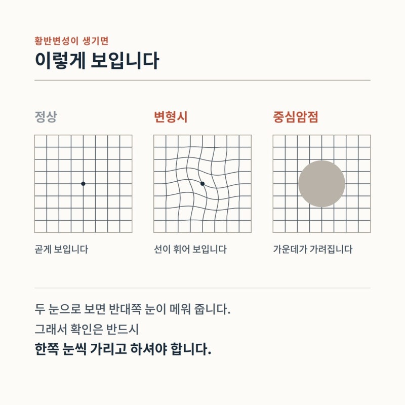
There are three representative symptoms:
① Metamorphopsia (Distorted Vision) — Straight lines look bent or wavy.
When fluid builds up under the macula or tissues are displaced upward, the retina becomes uneven and bumpy.
The image projected onto it is distorted as a result. Consequently, objects with naturally straight lines—such as door frames, bathroom tile lines, window mullions, utility poles, or white lines on pedestrian crossings—appear wavy or warped.
② Central Scotoma — The center of your vision appears blank, smudged, or grayed out.
When reading text, a letter in the middle seems erased; when looking at someone's face, the area around the nose and mouth appears hazy. Often, if you shift your gaze slightly to the side, it becomes clearer.
③ Decreased contrast and color perception, and micropsia/macropsia.
Reading in dim light becomes exceptionally difficult, and adjusting to bright environments takes much longer. Objects may also appear to be different sizes when comparing one eye to the other.
Here is the most critical takeaway:
We normally view the world with both eyes open.
So even if a defect arises in one eye, the healthy eye naturally compensates and covers for it.
In clinical practice, many patients who arrive saying, "I suddenly couldn't see yesterday," actually only noticed it by chance when covering one eye. The disease did not develop yesterday—it was just discovered yesterday.
This is why self-checks must always be performed covering one eye at a time.
3. How do you use the Amsler grid?
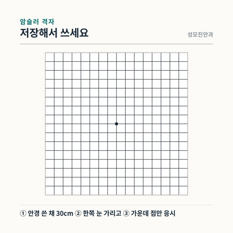
The Amsler grid is an image of a square containing a checkerboard pattern with a single dot in the center.
No special equipment is needed; you can save the image from this post and use it.
Here is the step-by-step process:
Wear your usual reading glasses or corrective lenses. If you have reading glasses, make sure to put them on.
Hold the grid approximately 30 cm away from your eyes in a well-lit room.
Completely cover one eye with the palm of your hand. Ensure no light leaks through your fingers.
With the uncovered eye, stare directly and steadily at the center dot. Do not move your gaze around the grid. This test evaluates how peripheral areas look while maintaining continuous fixation on the center.
Hold this view for 5 to 10 seconds, and observe:
Are any of the grid lines bent, distorted, or wavy?
Are there any areas that appear missing, blank, or blackened out?
Can you see all four corners of the grid?
Do any squares look unusually larger or smaller than the others?
Switch eyes and repeat the test exactly the same way.
How often should you do this?
If you have already been diagnosed with macular degeneration, or if you have wet AMD in one eye and need to monitor the other, it is recommended to check daily or every other day.
Even if you have no known conditions, building a habit of checking once a week allows you to detect subtle changes quickly.
A practical tip:
Remember or mark on paper how the grid looked the very first time you checked it.
In macular degeneration, what matters most is not just the fact that "it looks slightly wavy," but rather the progression: "it is wavier today than it was yesterday."
When to seek immediate emergency ophthalmic care:
▶ Lines that were straight yesterday suddenly appear distorted today
▶ A blank or darkened spot in the center starts expanding
▶ Vision in one eye suddenly turns blurry
▶ Objects suddenly look smaller (or larger) than normal
Wet macular degeneration can progress significantly within just a few days.
Saying, "I'll ask about it at my regular checkup next month," is the most dangerous postponement you can make.
4. Can you rely solely on the Amsler grid?

The straightforward answer is: no. We must be transparent about this.
Comprehensive studies have evaluated the diagnostic accuracy of the Amsler grid.
A systematic review and meta-analysis pooled 10 studies comprising 1,890 eyes (425 wet AMD eyes, 1,262 non-neovascular AMD eyes, and 203 normal eyes)
(Bjerager J et al., JAMA Ophthalmology, 2023).
Comparison Group | Sensitivity (Ability to detect disease) | Specificity (Ability to avoid false alarms) |
Compared to healthy eyes | 67% (95% CI 51–79%) | 99% (95% CI 85–100%) |
Detecting conversion to wet AMD in eyes with existing AMD | 71% (95% CI 60–80%) | 63% |
Here is how to interpret this table:
The first row looks reassuring, but it does not reflect the real clinical challenge.
What we truly need to catch is in the second row.
When using the Amsler grid to detect whether an eye with existing macular degeneration has transitioned into wet AMD, it misses roughly 3 out of 10 cases.
The authors concluded: "The real-world diagnostic performance falls short of the level typically recommended for monitoring the development of neovascular AMD."
Therefore, keep these two rules in mind:
▶ If you notice an abnormality on the Amsler grid → It is highly meaningful. Visit an eye clinic immediately.
▶ If the Amsler grid looks completely normal → It is not sufficient proof of safety. It can never replace regular fundus photography and OCT scans.
Self-testing is an interim bridge between clinical visits, not a replacement for them. If this distinction is missed, people often think, "The grid looks fine, so I must be okay," leading to dangerously delayed care.
5. Dry vs. Wet: What is the difference?

Macular degeneration is broadly divided into two forms.
They must be approached as if they are entirely separate conditions.
Classification | Dry (Non-exudative) | Wet (Exudative / Neovascular) |
Proportion | Majority of cases | Minority of cases |
Rate of Progression | Slow, over several years | Rapid (weeks to months), sometimes days |
Primary Findings | Drusen accumulation, pigmentary changes; geographic atrophy in late stages | Exudation and hemorrhage from choroidal neovascularization |
Onset of Symptoms | Gradual; late self-awareness | Sudden metamorphopsia and central scotoma |
Current Treatment | No established cure; focus on lifestyle modification and close observation | Intravitreal anti-VEGF injections |
Management Principle | Risk factor control + regular follow-up | Immediate clinical treatment |
In the aforementioned Korea National Health and Nutrition Examination Survey analysis, among the 2,222 macular degeneration cases, 2,085 were early-stage, 38 were geographic atrophy, and 99 were neovascular (wet) AMD
(Kim Y et al., Scientific Reports, 2025).
This means that the vast majority are still in the early stages—and that is good news.
It proves that for most people, timely management can still preserve their vision.
There is another important detail: wet AMD in Asian populations often presents differently than in Western populations.
A study of 321 Korean patients suspected of having exudative AMD at a tertiary hospital found that 24.6% (79 patients, 98 eyes) were diagnosed with Polypoidal Choroidal Vasculopathy (PCV)
(Byeon SH et al., Japanese Journal of Ophthalmology, 2008).
PCV can follow a different clinical course and respond differently to treatment compared to conventional wet AMD, often necessitating specialized testing like indocyanine green angiography (ICGA).
Under the single umbrella term "macular degeneration," distinct subtypes exist.
The overall number of domestic patients is also growing rapidly.
According to National Health Insurance data from 2013 to 2022, the annual prevalence of exudative AMD increased from 10.7 to 22.5 per 10,000 people, with patient counts surging from 27,273 to 127,044
(Kim MS et al., Journal of Korean Medical Science, 2025).
The same study projects that by 2040, prevalence will hit 46.2 per 10,000, totaling 374,282 patients.
6. What is my personal risk level?
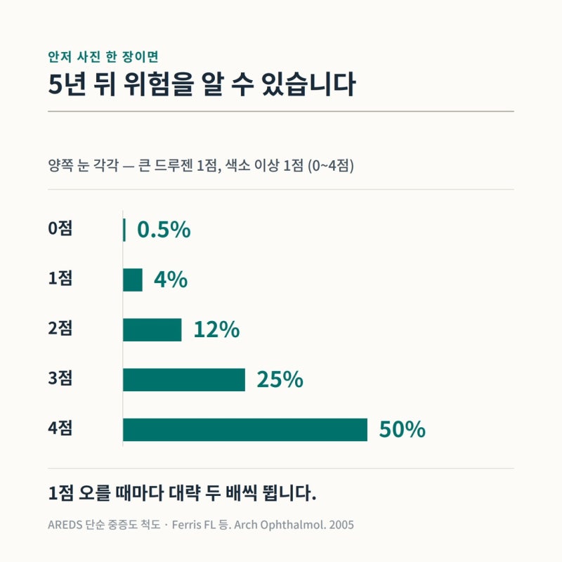
There is a validated scale that can estimate your future risk simply by analyzing fundus photographs.
This is the AREDS Simplified Severity Scale
(Ferris FL et al., AREDS Report No. 18, Archives of Ophthalmology, 2005).
Scores are calculated as follows:
For each eye:
① Presence of large drusen = 1 point
② Presence of pigment abnormalities = 1 point
Total score across both eyes ranges from 0 to 4 points.
Score | 5-Year Risk of Progression to Advanced AMD |
0 points | Approx. 0.5% |
1 point | Approx. 4% |
2 points | Approx. 12% |
3 points | Approx. 25% |
4 points | Approx. 50% |
Notice how the risk roughly doubles with each point: 12% → 25% → 50%.
This entire score can be determined from a single fundus photograph.
Even someone with zero visual symptoms can clearly know their exact risk score through one simple image.
Other well-established risk factors include:
▶ Age — The strongest risk factor. Risk rises steeply after age 50.
▶ Smoking — A meta-analysis showed that individuals with a history of smoking had a relative risk of 1.61 (95% CI 1.01–2.57) in cohort studies and 1.76 (95% CI 1.56–1.99) in case-control studies (Cong R et al., Annals of Epidemiology, 2008; 13 studies analyzed). Smoking cessation is the single most definitive modifiable risk factor for AMD.
▶ Family History — Having a first-degree relative with AMD significantly elevates your risk.
▶ Hypertension, cardiovascular disease, obesity, and intense UV exposure have also been associated with higher risk.
If your parents or siblings have macular degeneration, you should undergo a baseline fundus examination in your late 40s even without symptoms.
Having a baseline is essential to detect future changes early.
7. What tests are performed at the clinic?
① Visual Acuity & Refraction Testing — Establishes baseline best-corrected visual acuity.
② Fundus Examination — Performed after dilating the pupil (mydriasis) to evaluate the entire retina. Doctors examine drusen size, count, pigment changes, and check for bleeding or exudates. This is where the severity score is determined.
③ Optical Coherence Tomography (OCT) — The cornerstone of modern macular diagnosis. It scans cross-sections of the macula at micrometer resolution. It is non-contact, painless, and takes only a few seconds.
Subretinal fluid and retinal architectural disruption can be directly visualized.
OCT can catch fluid leakage at the earliest stages of wet AMD before the patient experiences any symptoms.
④ OCT Angiography (OCTA) — Visualizes retinal and choroidal microvasculature without dye injections to identify abnormal choroidal neovascularization.
⑤ Fluorescein Angiography (FA) — When indicated, contrast dye is injected intravenously to track leakage points. Indocyanine green angiography (ICGA) is particularly valuable for differentiating PCV.
Our clinic is equipped with advanced OCT and OCTA systems, allowing comprehensive evaluation combining fundus photography and high-resolution cross-sectional scans.
Testing notice: Pupillary dilation drops can cause light sensitivity and blurred near vision for about 4 to 6 hours.
Please avoid driving yourself to the appointment and bring sunglasses for comfort. Please also expect a short waiting period while your pupils fully dilate.
8. How is it treated? Can lost vision be restored?
There is currently no established medical cure to reverse dry AMD.
Treatment focuses on risk factor management, nutritional supplementation (Section 9 below), and periodic monitoring.
While this may sound passive, the primary goal of early monitoring is to catch the exact moment dry AMD transitions to wet AMD without delay.
The standard treatment for wet AMD is intravitreal anti-VEGF injection.
By blocking vascular endothelial growth factor (VEGF)—the chemical signal that stimulates abnormal vessel growth—it resolves leakage and hemorrhage.
Following local eye anesthesia, medication is injected directly into the vitreous cavity using an ultra-fine needle. The procedure itself is completed in seconds.
Typically, monthly loading doses are administered for the first few months, followed by interval adjustments based on treatment response.
However, several critical truths must be shared regarding this therapy:
First, this treatment controls the condition rather than curing it permanently. Most patients experience recurrences; thus, it is rarely a one-time treatment. It requires repeated injections and regular monitoring over several years. Discontinuing treatment can lead to rapid deterioration.
Second, damaged visual acuity rarely returns to normal. The primary goal of anti-VEGF therapy is to prevent further visual decline; vision improvement is a secondary bonus. Photoreceptors and nerve cells that have already atrophied or formed fibrotic scars cannot be regenerated.
Third, the timing of treatment initiation is decisive. In a US IRIS Registry study analyzing 162,902 eyes treated with anti-VEGF therapy, eyes with poorer vision at initial presentation gained more letters but ended up with significantly lower final vision (Ho AC et al., 2020; summarized in Phan LT et al., Clinical Ophthalmology, 2021). Where you start determines where you finish.
Fourth, we know the natural course of untreated disease. A systematic review of 2,293 untreated eyes with exudative AMD showed that visual acuity declined by approximately 15 ETDRS letters (about 3 lines on an eye chart) per year—reaching roughly a 30-letter loss at 2 years and a 45-letter loss at 3 years
(Elshout M et al., BMC Ophthalmology, 2017).
Fifth, injections carry potential risks. Common minor side effects include subconjunctival hemorrhage, temporary eye discomfort, and transient floaters. Rarely, endophthalmitis (intraocular infection), elevated intraocular pressure, retinal detachment, or retinal pigment epithelium tears can occur. Systemic risks such as stroke or myocardial infarction are also closely monitored. Injections must only be administered when clinically necessary based on definitive objective testing, not as a preventive measure.
Note: Even if significant vision loss has occurred, supportive options remain.
Low-vision aids—such as optical magnifiers, high-contrast task lighting, digital screen enlargers, and text-to-speech software—help restore daily independence.
Because macular degeneration spares peripheral vision, it does not cause total black-out blindness.
We emphasize this because unnecessary panic often causes excessive emotional distress.
9. Do supplements (Lutein / AREDS2) really help?
This is one of our most frequently asked questions.
The answer: "Yes, for specific individuals and within a specific range." Context is everything.
What is clinically proven: In the landmark AREDS trial involving 4,757 patients aged 55 to 80, a formulation of high-dose antioxidants and zinc reduced the risk of progression to advanced AMD by about 25% and visual loss by about 19%
(AREDS Report No. 8, Archives of Ophthalmology, 2001).
However, the key is who benefited: this preventive benefit was observed only in high-risk individuals—those with intermediate AMD in one or both eyes, or advanced AMD in one eye.
No clear benefit was found in individuals with no AMD or with only early-stage disease.
In other words, the evidence does not support taking these supplements preemptively "just because they're good for eye health."
The formulation was subsequently refined.
In the AREDS2 trial, beta-carotene was replaced with lutein and zeaxanthin. Over 10 years of follow-up (3,883 participants), the lutein/zeaxanthin group showed an additional 20% reduction in progression to advanced AMD compared to the beta-carotene group.
Furthermore, beta-carotene was found to nearly double the risk of lung cancer in former or current smokers
(Chew EY et al., AREDS2 Report 28, JAMA Ophthalmology, 2022).
Key points to remember:
Supplements are not preventive cures; they are supportive agents designed to slow disease progression in eyes that already have moderate degeneration.
Whether you should take them must be decided after determining your stage via a fundus examination. Starting them without an exam often leads to people who don't need them taking them, while those who genuinely need them miss out.
Current or former smokers must strictly avoid formulations containing beta-carotene. Always inspect ingredient labels carefully.
AREDS2 formulations contain high zinc levels. If you are taking other mineral multivitamins, check to ensure you are not exceeding safe intake limits.
Lutein does not improve eye strain or baseline refractive visual acuity. It specifically supports macular pigment optical density.
10. What lifestyle changes can lower my risk?
Listed in order of scientific evidence strength:
① Smoking Cessation: The most definitive intervention. Quitting smoking objectively lowers your risk. Age and genetics cannot be altered, but this can.
② Nutrition: Numerous observational studies suggest that diets rich in dark green leafy vegetables (spinach, kale) and oily fish rich in omega-3 fatty acids are beneficial. However, observational data cannot confirm absolute causation, and dietary intake is distinct from high-dose pill supplementation.
③ Managing Blood Pressure, Weight, and Blood Sugar: Cardiovascular risk factors significantly overlap with macular degeneration risks.
④ UV Protection: For extensive outdoor activities, wearing UV-blocking sunglasses and wide-brimmed hats is strongly advised.
⑤ Routine Fundus Examinations: While exams do not alter inherent biological risk, they are the only way to achieve early detection. This explains why over 95% of patients in epidemiological studies were completely unaware of their condition.
When visiting family during holidays:
Try asking your parents these three questions:
"Do you find it harder to read small newspaper print lately?"
"Do TV subtitles appear blurrier than before?"
"Can you cover one eye and look at that door frame for a second?"
It is surprisingly common for issues to be discovered with the third question.
Holidays offer a great opportunity to check the eye health of the entire family.
For information regarding pediatric vision and myopia management, please refer to our previous detailed post.
11. It might not be macular degeneration
Symptoms like "lines look crooked" or "the center looks blocked" can stem from other retinal disorders.
Because treatment strategies differ entirely depending on the underlying pathology, you should never self-diagnose.
▶ Macular Hole — A physical tear or hole forms in the center of the macula. It creates a distinct missing void in central vision and frequently requires vitreoretinal surgery.
▶ Epiretinal Membrane (Macular Pucker) — A delicate, fibrous membrane forms on the macular surface and contracts. It is a common cause of metamorphopsia and typically progresses very slowly.
▶ Central Serous Chorioretinopathy (CSC) — Commonly occurs in relatively young men aged 30 to 50. Associated with stress and corticosteroid use, many cases resolve spontaneously.
▶ Diabetic Macular Edema — If you have diabetes, this must be evaluated first.
▶ Retinal Vein Occlusion — Characterized by sudden, painless vision reduction.
▶ Optic Neuropathy or Migraine Aura — Flashing lights or zigzag geometric patterns that last 20 to 30 minutes before fading are often ocular migraine auras.
▶ Cataracts — Cause overall generalized blurriness, but do not make straight lines bend. Differentiating presbyopia from cataracts was covered in our separate article.
▶ Floaters & Flashes — Spots drifting in your field of view or lightning streaks originate from vitreoretinal traction, representing a distinct pathway from macular degeneration.
As a practical rule of thumb: distorted straight lines point to the macula; floating specks point to the vitreous; generalized cloudy haziness typically points to the cornea or lens.
An accurate diagnosis requires objective ophthalmic testing.
12. Frequently Asked Questions
Q. Does macular degeneration lead to total blindness?
The macula controls central vision, leaving peripheral vision preserved. While it can severely impair your ability to read or recognize faces, it rarely leads to total dark blindness where you cannot see your surroundings at all. Nevertheless, because its impact on quality of life is profound, early detection is essential. There is no need for excessive panic, but it should never be dismissed lightly.
Q. If I have macular degeneration in one eye, will it develop in the other?
The risk is definitely elevated. For patients with wet AMD in one eye, home self-monitoring and routine clinical exams of the fellow eye are paramount. Recall that an AREDS score of 4 points (large drusen and pigment abnormalities in both eyes) carries a roughly 50% 5-year progression risk.
Q. I was told I have drusen during a health checkup. Does that mean I have macular degeneration?
Drusen alone does not automatically equate to macular degeneration. Small drusen are common, normal aging findings. What matters clinically is the size, quantity, and whether pigmentary changes coexist. This combination determines your risk score. If you bring your screening report to the clinic, we can assess and explain your exact stage.
Q. I have been taking lutein for years. Should I continue?
It depends entirely on the status of your macula. If you have intermediate AMD or higher, it is an evidence-backed choice; if your macula is healthy or shows only minimal early changes, clinical benefit has not been demonstrated. A single fundus photo can answer this question clearly, so a quick evaluation is better than guessing. If you have a smoking history, make sure it is free of beta-carotene.
Q. How often should I get an eye exam?
Generally, adults over 50 with healthy eyes should be screened every 1 to 2 years. If drusen or pigmentary changes are present, every 6 to 12 months is recommended. If one eye has wet AMD, follow-ups are much more frequent. Exact intervals depend on individual clinical findings and should be tailored by your ophthalmologist.
Q. Does heavy smartphone or TV use cause macular degeneration?
There is currently no established scientific evidence showing that screen blue light causes macular degeneration. Prolonged screen use causes digital eye strain and dry eye disease, which are distinct pathologies from macular degeneration. Quitting smoking, managing blood pressure, and getting routine exams are far more important than screen time.
Q. Can I use the Amsler grid on a smartphone screen?
Yes, it is possible. However, ensure screen brightness is consistent and maintain an exact distance of about 30 cm every time to allow accurate comparison. Printing the grid on paper and sticking it on your refrigerator or bathroom mirror is a reliable way to make it a daily habit.
For more detailed visual guides on eye health, please visit St. Mary's Jin Eye Clinic's official YouTube channel, 'Jineye'.
In Conclusion
Please remember three essential takeaways:
First, always check one eye at a time. Both eyes open will mask defects in a single eye.
Second, a normal Amsler grid does not guarantee complete safety. When screening for active wet AMD, the grid misses about 3 out of 10 cases. Self-testing complements regular screening exams—it does not replace them.
Third, if anything looks different from yesterday, do not wait. With wet AMD, your starting point dictates your finishing vision.
Macular degeneration is detected in the early stages in most cases, and early detection offers a wide array of protective interventions.
Taking a single fundus photograph when you have no symptoms is the most affordable, definitive investment you can make for your vision today.
Please feel free to reach out if you have any questions.
St. Mary's Jin Eye Clinic
337 Gangnam-daero, Seocho-gu, Seoul (1329-10 Seocho-dong)
5-minute walk from Gangnam Station Exit 5 · Free parking available in the building
Clinic Hours
Weekdays: 09:00 – 18:00 (Lunch: 13:00 – 14:00)
Saturdays: 09:00 – 15:00 (No lunch break)
Closed on Sundays and Public Holidays
Phone: 02-577-7782
※ This article is intended for general informational purposes only and does not substitute for personalized medical diagnosis or treatment. The Amsler grid referenced here is a supplementary self-monitoring aid, not a definitive diagnostic test. Cited statistics reflect the findings of their referenced study cohorts; individual outcomes may vary based on ocular health, systemic conditions, and medications. All treatments carry limitations and potential side effects; diagnostic and treatment decisions must always be made in consultation with an eye care specialist.
[References]
Kim Y, Han K, Kim JH. Prevalence and risk factors of undiagnosed age-related macular degeneration: the Korea National Health and Nutrition Examination Survey 2017–2020. Scientific Reports. 2025;15:10444
Bjerager J, Schneider M, Potapenko I, et al. Diagnostic Accuracy of the Amsler Grid Test for Detecting Neovascular Age-Related Macular Degeneration: A Systematic Review and Meta-analysis. JAMA Ophthalmology. 2023;141(4):315-323
Ferris FL, Davis MD, Clemons TE, et al. A simplified severity scale for age-related macular degeneration: AREDS Report No. 18. Archives of Ophthalmology. 2005;123(11):1570-1574
Age-Related Eye Disease Study Research Group. A randomized, placebo-controlled, clinical trial of high-dose supplementation with vitamins C and E, beta carotene, and zinc for age-related macular degeneration and vision loss: AREDS Report No. 8. Archives of Ophthalmology. 2001;119(10):1417-1436
Chew EY, Clemons TE, Agrón E, et al. Long-term Outcomes of Adding Lutein/Zeaxanthin and ω-3 Fatty Acids to the AREDS Supplements on Age-Related Macular Degeneration Progression: AREDS2 Report 28. JAMA Ophthalmology. 2022;140(7):692-698
Cong R, Zhou B, Sun Q, Gu H, Tang N, Wang B. Smoking and the risk of age-related macular degeneration: a meta-analysis. Annals of Epidemiology. 2008;18(8):647-656
Elshout M, van der Reis MI, de Jong-Hesse Y, Webers CAB, Schouten JSAG. Tracing the natural course of visual acuity and quality of life in neovascular age-related macular degeneration: a systematic review and quality of life study. BMC Ophthalmology. 2017;17:120
Byeon SH, Lee SC, Oh HS, Kim SS, Koh HJ, Kwon OW. Incidence and clinical patterns of polypoidal choroidal vasculopathy in Korean patients. Japanese Journal of Ophthalmology. 2008;52(1):57-62
Kim MS, Nam S, Park SJ, Lee J, Woo SJ. 10-Year Change and Projection in Prevalence and Incidence of Exudative Age-Related Macular Degeneration in Korea. Journal of Korean Medical Science. 2025
Phan LT, Broadhead GK, Hong TH, Chang AA. Predictors of Visual Acuity After Treatment of Neovascular Age-Related Macular Degeneration — Current Perspectives. Clinical Ophthalmology. 2021;15:3351-3367
American Academy of Ophthalmology. Age-Related Macular Degeneration Preferred Practice Pattern. Ophthalmology. 2025;132(2)