2026-09-17
Floaters in Your Vision? Harmless Eye Floaters vs. Warning Signs Requiring Immediate Eye Care
Floaters are usually a normal sign of aging, but sudden floaters accompanied by flashes or vision changes may indicate a retinal tear or detachment requiring immediate dilated eye examination.


If you suddenly notice dark spots or thread-like shapes drifting across your field of vision—and they seem to move whenever you move your eyes—you are experiencing eye floaters (myodesopsia).
To put it simply, most floaters are a natural consequence of aging and do not require treatment.
However, among 100 patients with sudden-onset floaters, about 14 are found to have an accompanying retinal tear. If left untreated, this can progress to retinal detachment, a serious condition that can lead to permanent vision loss.
Because it is impossible to distinguish between "benign floaters" and "dangerous floaters" based on symptoms alone, a dilated fundus examination is the only definitive way to directly assess the retina.
Hello, this is St. Mary Jin Eye Clinic.
Today, we will provide an evidence-based overview of why floaters develop, which warning signs warrant immediate attention, and what eye specialists look for during an examination.
1. What exactly do floaters look like?

Different people describe them in different ways.
Gnats, black specks, strands of thread, hairs, cobwebs, heat hazes, or translucent smudges—they all share these common features:
▶ They follow your gaze when you move your eyes, drifting slightly after your eyes stop.
▶ They become much clearer when looking at a bright sky, a white wall, or a blank piece of paper.
▶ When you try to focus on them directly, they seem to drift away.
This occurs when opacities within the vitreous humor—the clear gel filling the eye—cast shadows onto the retina.
In other words, what you see is not an insect outside your eye, but a shadow cast inside it.
2. Why do they occur? — The Vitreous and Posterior Vitreous Detachment
The inside of the eye is filled with a transparent gel called the vitreous humor.
In our youth, it has an elastic, egg-white-like consistency firmly attached to the retina. As we age, however, it gradually liquefies and shrinks in volume.
Eventually, the contracted vitreous completely separates and peels away from the retina.
This event is known as a Posterior Vitreous Detachment (PVD), a natural aging process that typically occurs between the ages of 50 and 70.
Because the separated boundary creates clumps that cast shadows, floaters often suddenly appear or dramatically increase during this period.
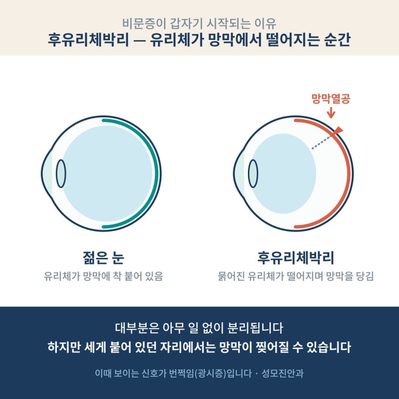
The critical issue lies in how the vitreous separates.
In areas where the vitreous is unusually firmly adherent to the retina:
▶ If it pulls and mechanically stimulates the retina → Flashes of light (photopsia) appear in peripheral vision, even with eyes closed.
▶ If the traction tears the retina → A retinal tear occurs.
For most people, the separation happens cleanly without incident. In some individuals, however, tears do form—and identifying these tears is the central objective of a clinical evaluation for floaters.
3. 14 out of 100 — The Likelihood of Dangerous Floaters
The common belief that "floaters are harmless" is only half true. Let us look at the clinical data.

In a meta-analysis of 17 studies covering over 20,000 patients presenting with acute-onset floaters or flashes:
▶ Retinal tears were diagnosed in approximately 14% of patients.
▶ When accompanied by hazy vision or obscured sight (vitreous hemorrhage), the risk of a tear increased dramatically (likelihood ratio ~10).
▶ Subjective vision loss was also a significant warning sign (likelihood ratio ~5).
(Hollands H et al., JAMA, 2009)
There is another critical finding to keep in mind.
Even when an initial dilated exam is completely normal, around 3% of patients develop a new retinal tear within the following few weeks.
This underscores why it is essential to return for a repeat examination if new symptoms arise, even after an initial clear checkup.
(Nature Eye, 2024 — Prospective study of community referrals for PVD)
In short: most patients with floaters have benign cases.
However, no one can tell whether you are in the clear without directly inspecting the retina.
The intensity of visual symptoms does not always correlate with the severity of underlying pathology.
4. 4 Warning Signs Requiring Immediate Eye Care
If you experience any of the following symptoms, do not delay—visit an eye clinic either the same day or within a few days.

① A sudden, dramatic increase in the number of floaters: Seeing one or two floaters multiply into dozens, or seeing what looks like a shower of black pepper. This can indicate bleeding from a torn retinal blood vessel.
② Flashes of light (photopsia): Seeing lightning streaks or camera-like flashes in your peripheral vision, especially in the dark or with your eyes closed. This indicates vitreous traction on the retina.
③ A curtain-like shadow over your field of vision: A dark veil or curtain creeping in from the periphery. This is a medical emergency that indicates a retinal detachment may have already started.
④ A sudden drop in visual acuity: Blurry vision, distorted lines, or a darkening in the center of your vision in one eye.
Think of ① and ② as "signs requiring prompt evaluation," and ③ and ④ as "emergency-level signs."
Furthermore, regardless of symptoms, individuals with high myopia must take floaters very seriously.
Myopic eyes have an elongated axial length, stretching the retina thin and making it significantly more prone to tears during posterior vitreous detachment. We discussed the relationship between myopia and retinal detachment risk in detail in our previous post.
https://blog.naver.com/jineye-center/224386510176
5. What happens if a retinal tear is found?
This is the fundamental reason why floaters require medical evaluation.
The treatment approach differs completely depending on whether the issue is caught at the tear stage or after it has progressed to detachment.
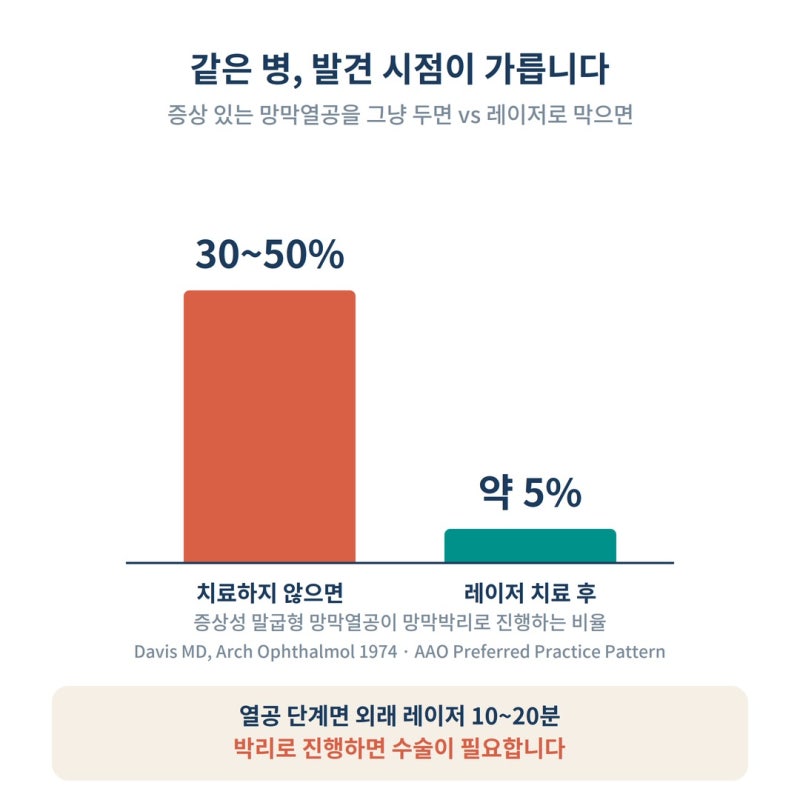
At the retinal tear stage — The tear can be sealed in an outpatient setting using retinal laser photocoagulation, effectively "welding" the surrounding tissue down.
It requires no anesthetic injections, no hospital admission, and takes only 10 to 20 minutes.
While an untreated, symptomatic horseshoe retinal tear progresses to retinal detachment in 30% to 50% of cases, timely laser treatment reduces this risk down to roughly 5%.
(Davis MD, Arch Ophthalmol, 1974 / American Academy of Ophthalmology PPP)
If it progresses to retinal detachment — Outpatient laser alone is no longer viable; surgical intervention (such as vitrectomy or scleral buckling) becomes necessary.
If surgery is delayed and the detachment involves the macula (the center of clear vision), vision may not fully recover even after an anatomically successful operation.
Two different outcomes of the exact same disease course: a 20-minute outpatient laser procedure versus major vitreoretinal surgery, separated by just a matter of days.
This stark reality illustrates why floaters should never simply be dismissed with "it's probably fine."
6. What tests are performed at the eye clinic?
The cornerstone of a floater evaluation is the dilated fundus examination.
Mydriatic drops are administered to enlarge the pupils. After waiting 15 to 20 minutes, the ophthalmologist thoroughly inspects the entire retina through the dilated pupil—paying special attention to the peripheral retina where tears most frequently occur.
When necessary, scleral depression (gentle pressure applied to the outside of the eye) is used to bring the extreme periphery into view.
Because tears are tiny and often hidden along the far edges, they can be missed on undilated eye exams or standard fundus photography.
In fact, studies have reported that a significant proportion of retinal tears cannot be detected using a standard slit-lamp exam alone. This is why pupil dilation, though slightly inconvenient, cannot be skipped.
Precautions on the Day of the Exam
▶ Your vision will be sensitive to glare, and near vision will remain blurred for 4 to 6 hours after dilation.
▶ Do not drive yourself; use public transportation or have a companion accompany you.
▶ Bringing sunglasses will make your trip home much more comfortable.
Even if the initial exam is normal, remember that while PVD is actively progressing, any new symptoms (increased flashes, a sudden surge in floaters, curtain-like shadows) warrant immediate re-examination.
7. Frequently Asked Questions
Q. Can floaters be eliminated through treatment?
Simple floaters without any underlying retinal disease are generally not targeted for treatment. Over time, the opacities tend to settle downward, or the brain adapts to them through neuroadaptation, making them much less noticeable. Although surgical removal (vitrectomy) or laser vitreolysis exists, the potential risks typically outweigh the benefits; therefore, they are considered only in rare, extreme cases where floaters severely impair daily life.
Q. Should I always visit an eye clinic if I have floaters?
Any floater that is "sudden in onset or has changed abruptly" definitely requires at least one dilated fundus examination. Long-standing floaters that have remained unchanged for years are not an emergency. However, if their number increases or if flashes accompany them, even long-standing floaters need to be re-evaluated.
Q. I only see flashes of light (photopsia) without any floaters. Do I still need an exam?
Yes, isolated photopsia is still an indication for an exam. It is a direct signal that the vitreous gel is tugging on the retina. As an aside, if you see shimmering zigzag lines that spread over 10 to 30 minutes, disappear, and are followed by a headache, it could be an ocular migraine rather than an eye problem; an eye exam is the safest way to differentiate between the two.
Q. Can young people also get floaters?
Yes, they can. High myopia in particular accelerates vitreous liquefaction, making it possible for floaters and posterior vitreous detachment to develop in people in their 20s and 30s. Sudden-onset floaters in young, highly myopic patients require the exact same thorough examination as in older adults.
Q. If PVD has occurred in one eye, what about the other eye?
It is very common for the fellow eye to undergo the same process within months to a few years. If new floaters or flashes begin in the opposite eye, you should undergo the same dilated examination.
Q. Can I rest easy if the test results are normal?
A normal result means your retina is healthy at that specific point in time. However, until the posterior vitreous detachment process is fully complete, there is a small chance (around 3%) of developing a new tear. Just keep in mind the fundamental rule: return for a re-check if new symptoms appear.
For more detailed information on eye health, please visit the St. Mary Jin Eye Clinic YouTube channel, 'Jjin Eye Clinic'.
In Conclusion
The vast majority of floaters are completely harmless.
Yet there is no reliable way to confirm whether they are harmless based solely on subjective feelings, and confirming their safety takes just one dilated fundus examination.
If floaters have suddenly appeared, multiplied rapidly, or are accompanied by flashes of light—do not put it off; have them checked.
When caught at the retinal tear stage, the condition can be resolved with a 20-minute outpatient laser session; left unattended, it can turn into major surgery.
Please feel free to reach out if you have any questions.
St. Mary Jin Eye Clinic
337 Gangnam-daero, Seocho-gu, Seoul (1329-10 Seocho-dong)
5-minute walk from Gangnam Station Exit 5 · Free parking available on site
Clinic Hours: Weekdays 09:00 - 18:00 (Lunch break 13:00 - 14:00)
Saturdays 09:00 - 15:00 (No lunch break)
Closed on Sundays and Public Holidays
Tel: 02-577-7782
※ This article is intended for general medical informational purposes only and does not substitute for personalized medical diagnosis or treatment. Recommended actions may vary depending on individual symptoms and clinical findings; please consult a board-certified ophthalmologist.
[References]
Hollands H et al. Acute-onset floaters and flashes: is this patient at risk for retinal detachment? JAMA. 2009
Posterior vitreous detachment and retinal tear — a prospective study of community referrals. Eye (Nature). 2024
Davis MD. Natural history of retinal breaks without detachment. Arch Ophthalmol. 1974
American Academy of Ophthalmology. Posterior Vitreous Detachment, Retinal Breaks, and Lattice Degeneration Preferred Practice Pattern
EyeWiki (AAO). Posterior Vitreous Detachment